


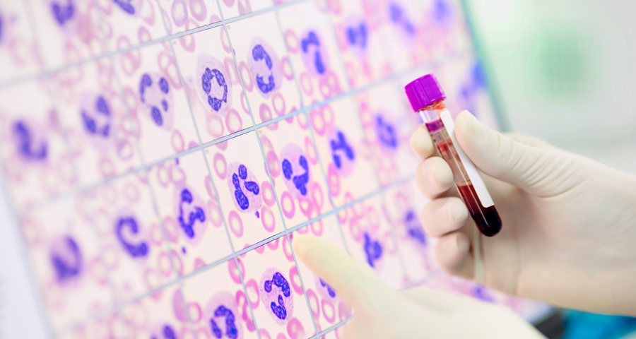
The quantity of each type of cell in a patient’s blood is determined using hematology analyzers. The role and significance of each blood component have just lately been realized. As blood circulates throughout the body, it nourishes each cell while also removing waste. There is no change in the body that does not result in a change in the blood, according to theory. More markers for specific occurrences are being sought by hematologists.
Functioning of Hematology Analyzers
- Measurement of RBC
- RF/DC Detection Method:
Changes in direct-current resistance detect the size of Hematology stained blood cells, while changes in radio-frequency resistance detect the density of the blood cell interior. A blood sample is aspirated and analyzed before being diluted to the appropriate ratio and sent to the appropriate detection chamber. Changes in direct-current resistance detect the size of the blood cell, and changes in radio0frequency resistance detect the density of the blood cell interior (size of the nucleus), both of which are sensed as electrical pulses.
- Hydro-Dynamic Focusing (DC Detection):
The sample nozzle is located in front of the aperture and in the center of the detector. After being driven into the conical chamber from the sample nozzle, the dilute sample is surrounded by a front sheath reagent and passes through the aperture center. The diluted sample of Hematology stains is enclosed by the back sheath reagent and transferred to the catcher tube after passing through the aperture. This avoids the drifting back of blood cells in this location, as well as the creation of spurious platelet pulses. This approach enhances the precision and repeatability of blood counts. Because the blood flows in a straight path via the aperture, irregular blood call pulses are avoided.
- Flow Cytometry Method Using Semiconductor Laser:
Physiological and chemical features of cells and other biological particles are studied using cytometry. Hematology stains, cells, and particles are analyzed using flow cytometry as they pass through extremely tiny flows. Aspirate a blood sample, measure it, dilute it to the necessary ratio, then stain it. After that, the sample is put into the flow cell. To the blood cells traveling through the flow cell, a semiconductor laser beam is emitted. The photodiode receives forward scattered light, whereas the photomultiplier tube receives laterally scattered light and lateral fluorescent light. This light is transformed into electrical pulses, allowing blood cell information to be obtained.
- Forward Scattered light and lateral Scattered Light:
When there are barriers in light, such as particles, the light beam scatters in different directions from each obstruction (light scattering). It is possible to gather information on cell size and material qualities by detecting scattered light.
Light scattering happens when a laser beam is directed upon Hematology stains and blood cell particles. The strength of light scatter is affected by a variety of parameters, including particle dimension and viewing angle. The size of blood cells is determined by forward scattered light, whereas the inside of the cell is determined by laterally scattered light (nucleus).
- Lateral Florescent light:
When light is shone over fluorescent material, such as stained blood cells or Hematology stains, it produces light with a longer wavelength than the original light. As the stain concentration rises, the intensity of the fluorescent light rises with it. The degree of blood staining may be determined by measuring the intensity of the light emitted. The XE2100 detects fluorescent light that is emitted sideways. Fluorescence light is emitted in all directions.
- Measurement of WBC
- WBC/BASO channel
STROMATOLYSER-FB, an acid haemolytic reagent, is used to lyse RBCs. The degranulation of Basophils is specifically suppressed by this reagent, resulting in their isolation from other WBC. Following this reaction, the sample is evaluated using flow cytometry, which uses a semiconductor laser to collect forward and side scattered light information, resulting in a WBC/BASO scattergram. WBC and Basophil counts are obtained by studying this scattergram.
- WBC 4-part differential
STROMATOLYSER-4DL is used for RBCs. The reagent also works on the WBC membrane, allowing dye to flow through. The dye STROMATOLYSER-4DS (dying solution) is then added, allowing the dye to enter the WBC at the damaged section of its membrane and stain the DNA and RNA therein. Following this reaction, a flow cytometry analysis of the sample of Hematology stains is performed, utilizing the s semiconductor laser to detect forward and side scattered light information, and a 4-DIFF scattergram is created. 4-parameter counts (lymphocytes, monocytes, Eosinophils, Neutrophils, and Basophils) are obtained by studying this scattergram.
Conclusion
Hematology Analyzers’ primary role and operation are to The cells are passed through an aperture one at a time utilizing hydrodynamic focusing. A laser is aimed at them during this process, and the dispersed light is measured from numerous angles. The absorbance is also taken into account. The intensity of the dispersed light and the amount of absorption can be used to identify the cell.